Pulmonic Stenosis in Dogs
Home » Pulmonic Stenosis in Dogs
What is Pulmonic Stenosis (PS)?
- Pulmonic Stenosis is one of the most commonly reported congenital heart defects in dogs, yet rarely reported in cats.
- It can occur in any breed, but is seen more frequently in Chihuahuas, Boxers, Labradors, Newfoundlands, West Highland White terriers, with known patterns of inheritance in both Beagles and English Bulldogs.
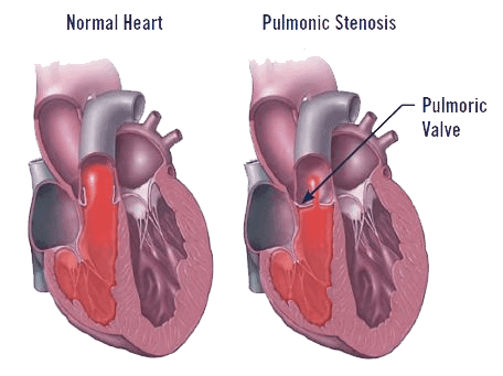
- Pulmonic stenosis is a defect involving the pulmonic valve and/or the surrounding structures of the right ventricle and pulmonary artery. The pulmonic valve normally opens during ventricular contraction allowing deoxygenated blood to be pumped from the right ventricle to the lungs. PS is a narrowing of this region (stenosis) resulting in an increased workload on the right ventricle causing hypertrophy (excessive heart muscle thickening).
- The most common form of the defect involves thickened, immobile pulmonic valve leaflets, however some forms of this disease are more complicated. One such defect involving an aberrant right coronary artery in both English Bulldogs and Boxers is important to recognize, as this form of PS is not as amenable to the most common interventional procedures.
How is it diagnosed?
- Dogs with PS rarely show symptoms as puppies.
- A murmur may be noted during a puppy’s first veterinary visit prompting the need for further investigation.
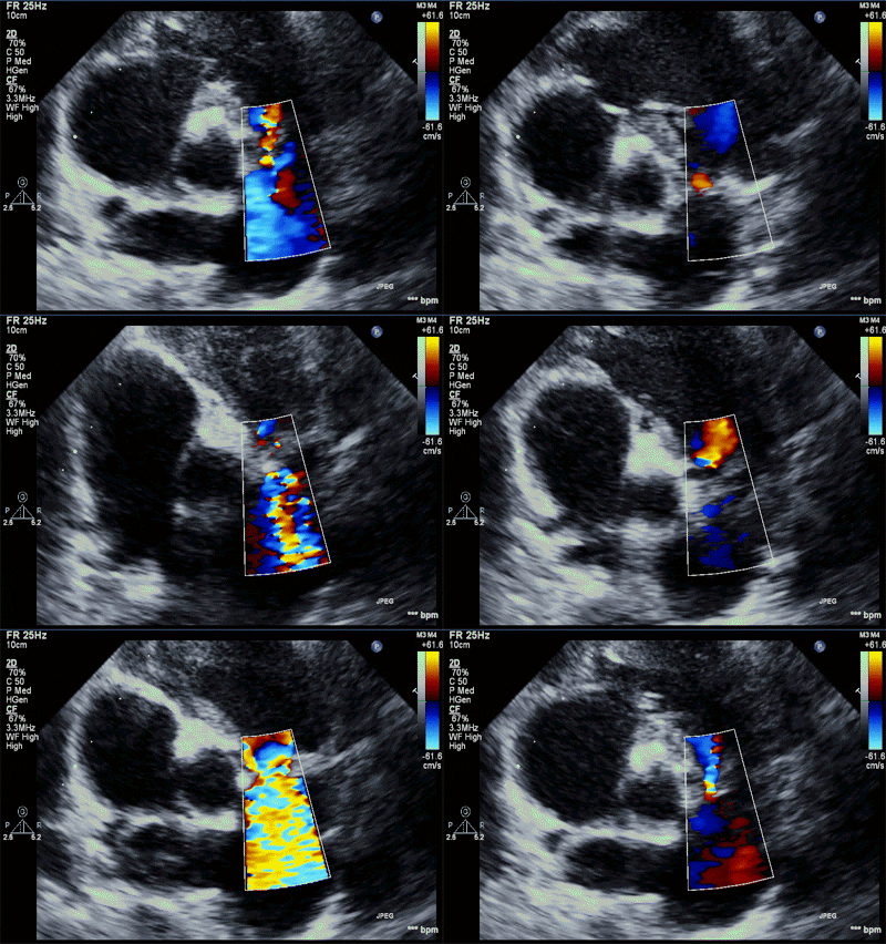
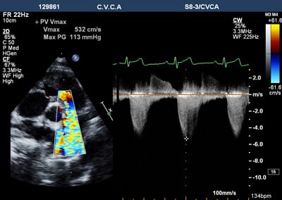
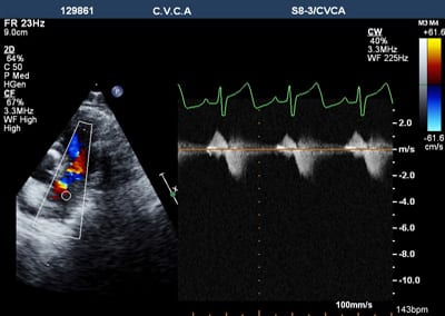
- A presumptive diagnosis can be made by your family veterinarian based on breed, physical exam findings and chest radiographs, however, a definitive diagnosis requires an echocardiogram by a veterinary cardiologist.
- An echocardiogram provides important information on the severity of the defect, detects any concurrent cardiac abnormalities that may complicate medical or surgical intervention, and helps to guide treatment.
Severity of Pulmonic Stenosis
- Mild PS: Pets usually live normal lives usually without the need for medications.
- Moderate to severe PS may result in:
- exercise intolerance
- shortness of breath
- fainting
- fatal arrhythmias (rare)
- congestive heart failure (rare) most commonly seen as abdominal effusion (ascites)
How is it treated?
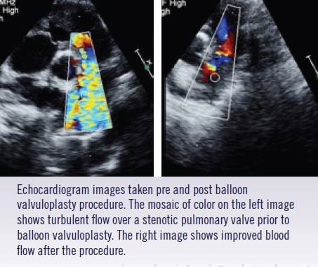
- For moderate to severe disease the treatment of choice is usually balloon valvuloplasty.
- Minimally invasive catheterization procedure by which a balloon is guided by fluoroscopy across the area of stenosis and inflated under pressure, resulting in stretching or tearing open of the abnormal valve, improving blood flow.
- In patients with severe disease, this often alleviates clinical signs, improves quality of life and prolongs survival.
- Medical therapy may also be recommended to decrease the incidence of arrhythmias or in cases where congestive heart failure is already present.
Our medical staff will be happy to answer any questions you may have or discuss treatment options in more detail. Our goal is an open collaboration with your veterinarian to develop an individualized treatment plan that will provide optimal quality care for your canine family member.
